
References
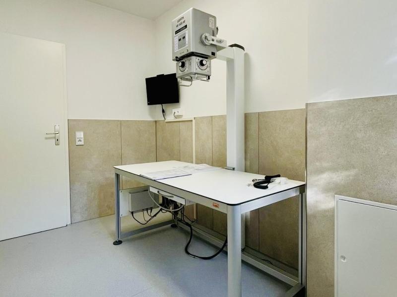
Stationary X-ray system for the small animal practice Grüne Aue
On February 4th, 2025, we were delighted to deliver, assemble, install, and train the new complete X-ray system for the small animal practice Grüne Aue in Zeitz. Dr. Hofmann and Dr. Schulze opted for our GIERTH combiVet S X-ray table with the GIERTH HF 400M X-ray machine and a stationary digital system from CANON (CXDI-702C). Both we and the owners were very satisfied with the service, and we are delighted to have happy customers.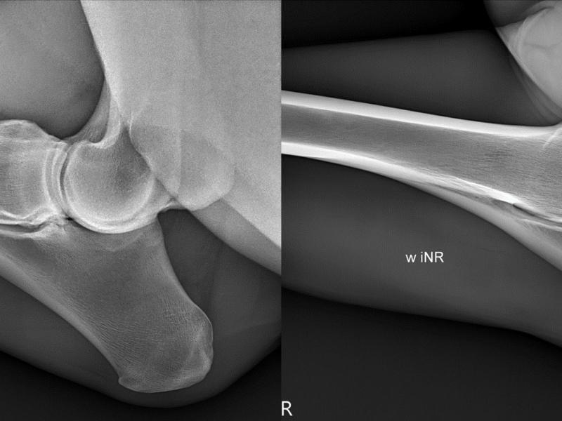
First installation of AI-based (Intelligent Noise Reduction) software at the equine clinic in Leichlingen
At the end of last year, GIERTH X-Ray international GmbH offered the equine clinic in Leichlingen the option of upgrading their X-ray technology, which they had been using successfully for many years with the detector solution from CANON Medical Components.
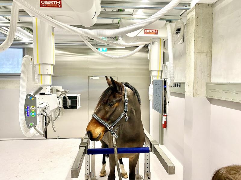
World's first multi-telescope Equine Radiography and Fluoroscopy Diagnostic Imaging System in Zurich
GIERTH X-Ray international is very proud to announce that with our partners Control-X Medical, CANON Medical Components Europe and CPI we have successfully installed the world’s first multi-telescope Equine Radiography and Fluoroscopy Diagnostic Imaging System at the Vetsuisse Faculty of the University of Zürich; one of the premier veterinary universities in Europe.

New Dual Telescope Veterinary Radiographic System at Medizinisches Pferdezentrum am Niederrhein
Another successful installation by GIERTH X-Ray international GmbH was realised in April 2022 at Medizinisches Pferdezentrum am Niederrhein in Straelen.
Al Wabra Wildlife Preservation
Al Wabra Wildlife Preservation is a private wildlife collection powered by Sheikh Saoud Bin Mohammed Bin Ali Al Thani's passion for nature.
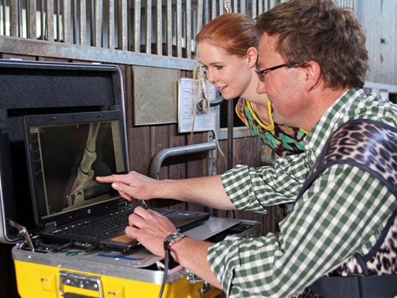
Dr. med. vet. Claudius Krieg - Equine veterinary specialist
Since January 2011, Dr. Claudius Krieg, specialist in orthopedic equine medicine, is working together with his two assistants in a mobile equine clinic dedicated to provide your horse with the best treatment.
© Copyright 2026 GIERTH X-Ray international GmbH | Riesa
We will be pleased to advise you:
+49 (0) 35 25 - 51 24 59
GIERTH X-Ray international GmbH
Am Südspeicher 4
D-01587 Riesa
Germany
Telefon: +49 (0) 35 25 - 51 24 59
Telefax: +49 (0) 35 25 - 51 24 24
E-Mail: info@gierth-x-ray.de
Medical Partner:
ATOMED X-Ray GmbH
![]()